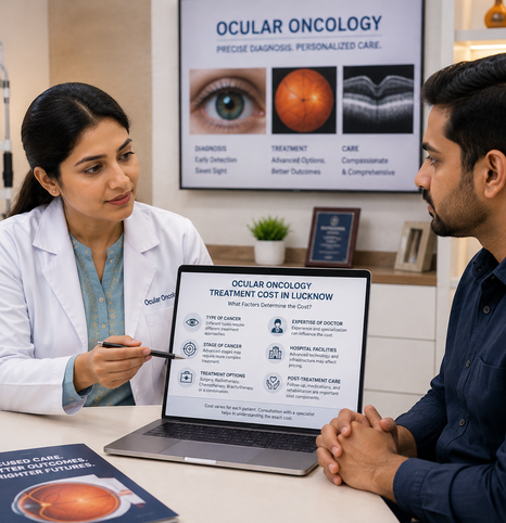
Ocular oncology focuses on the early detection, diagnosis, and treatment of eye tumours affecting the eye and surrounding structures.
Ocular oncology is a specialised branch of ophthalmology that focuses on eye tumours and related conditions. It includes diagnosis and management of both benign and malignant growths.
Intraocular layers including the retina, choroid, and ciliary body.
Upper and lower lid tissues, lid margins, and sebaceous glands.
Bony socket, orbital muscles, connective tissues, and nerves.
Clear mucous membrane covering the white of the eye and inner lids.
Lacrimal drainage pathways, tear glands, and adjacent facial tissues.
Some tumours may cause noticeable symptoms, while others can develop without pain or obvious warning signs. Early diagnosis helps doctors understand the clinical nature of the condition and guide appropriate treatment.
Determining whether the growth is benign, pre-cancerous, or malignant.
Measuring precise basal diameter, thickness, and elevation with imaging.
Assessing proximity to the macula, optic nerve, or sensitive ocular margins.
Ocular growths vary significantly in their origin, behavior, and required management. Explore the common conditions evaluated in ocular oncology.
Retinoblastoma is the most common primary intraocular cancer in infants and young children. It develops in the cells of the retina and often presents with a white pupil reflection (leukocoria) or strabismus.
Ocular melanoma is the most frequent primary intraocular tumour in adults, usually arising in the uveal tract (choroid, ciliary body, or iris). Specialized diagnostic imaging is crucial for accurate staging.
Growths on the eyelids range from benign cysts and papillomas to malignant lesions like basal cell or sebaceous carcinoma. Accurate diagnosis helps ensure clear margins while preserving eyelid structure.
Orbital tumours develop within the eye socket behind or around the eyeball. They can cause progressive proptosis (protrusion of the eye), double vision, or pressure around the orbital rim.
Conjunctival tumours arise on the surface of the eye. They include benign naevi, ocular surface squamous neoplasia (OSSN), and conjunctival melanomas, requiring careful biomicroscopic inspection.
Metastatic eye tumours occur when cancer cells spread to the uvea or orbit from primary sites elsewhere in the body. Coordinated multi-specialist care is vital.
Many ocular growths, such as choroidal naevi, haemangiomas, and benign cysts, are non-cancerous. Structured periodic follow-up is necessary to rule out malignant transformation.
Treatment planning depends on tumour type, size, location, and clinical stage. Ocular oncology combines detailed eye examinations, imaging, laboratory tests, and specialist care.
Book Diagnostic WorkupDetailed eye examination including slit-lamp biomicroscopy and dilated indirect ophthalmoscopy.
Imaging / diagnostic assessment with high-resolution ocular ultrasound and DICOM scans.
Required laboratory assessment and tissue pathology when clinically indicated.
Specialist evaluation and treatment planning tailored to each patient's condition.
The right ocular oncology treatment depends on the patient's condition. Doctors consider tumour type, size, location, and clinical stage when recommending the most appropriate pathway.
Some small or benign tumours may not require immediate surgery. The doctor may recommend regular observation and follow-up examinations to monitor changes over time and guide timely care.
Approximate cost for consultation and investigations. Actual cost depends on individual evaluation.
Schedule EvaluationCertain eye lesions and selected tumours may be treated with laser photocoagulation, transpupillary thermotherapy, or other local therapies. Suitability depends on tumour type and anatomical location.
Per session approximate cost. Actual treatment requirements and cost depend on clinical evaluation.
Inquire About Laser CareSome eye cancers may require chemotherapy or other drug-based treatments. Retinoblastoma and certain metastatic tumours are examples where systemic or local drug treatment may be considered based on the patient's age, tumour type, and overall health.
Approximate range depending on drug protocol, cycles, and specialist supervision.
Consult Oncology TeamSurgery may be recommended for certain tumours affecting the eyelid, orbit, conjunctiva, or other eye structures. The surgical approach depends on size, location, and nature of tumour. In some cases, tissue is sent for laboratory examination to confirm diagnosis.
Approximate source range. Actual cost varies with procedure complexity and tissue reconstruction.
Request Surgical OpinionFor selected eye tumours, doctors may consider eye-conserving treatments such as plaque therapy or a combination of local and systemic treatments. The goal is to control the tumour while preserving the eye and useful vision whenever possible.
Approximate cost according to personalized eye-conserving treatment plan.
Discuss Eye PreservationTreatment cost varies for every patient depending on diagnosis, complexity, and individual clinical requirements.
Ocular oncology requires trained clinical expertise, precise diagnostic infrastructure, and clear patient guidance. Look for these essential benchmarks:
Schedule A ConsultationSpecialists with focused training in identifying and managing benign and malignant eye growths.
Access to standardized digital imaging and pathology reporting to confirm tissue diagnosis.
Competence across both childhood conditions (like retinoblastoma) and adult intraocular growths.
Seamless collaboration involving oncology and radiology when systemic management is required.
Clear communication regarding findings, treatment choices, and realistic clinical expectations.
Clear financial estimates for investigations, surgical procedures, and ongoing follow-up.
Scheduled surveillance protocols to monitor eye health and check for recurrence.
Some ocular oncology treatments may be covered by health insurance. We encourage patients to review their policy terms for peace of mind.
A structured clinical pathway ensures thorough evaluation, informed decision-making, and emotional reassurance throughout your care.
Detailed discussion of medical history, eye symptoms, and preliminary clinical examination.
Targeted scans and ophthalmic investigations (typically 60–90 minutes).
Specialist review of all findings to confirm tumour type, dimensions, and anatomical boundaries.
Formulating a tailored care plan (observation, laser, surgery, or medication) with emotional counseling support.
Periodic follow-up examinations to ensure long-term stability and recurrence monitoring.
If you or your family member notice any of the following signs, seek professional ophthalmic assessment promptly:
Timely clinical evaluation provides clarity and helps guide the safest next steps.
Book Professional EvaluationKlarity Eye Care offers specialised eye care for patients who need evaluation for ocular tumours. The approach focuses on detailed diagnosis, appropriate treatment planning, and regular follow-up.
Modern diagnostic technology helps specialists examine eye structures and assess unusual growths. Whether the patient is a child or an adult, early evaluation can help guide timely care.
If you are concerned about an unusual growth, change in vision, persistent swelling, or have been referred for tumour assessment, seek appropriate professional evaluation.
Speak with our ocular oncology coordination desk.
Thank you for contacting Klarity Eye Care Hospital. Our ocular oncology care coordinator will connect with you shortly to schedule your evaluation.