Glaucoma Treatment in Lucknow
Glaucoma treatment focuses on controlling eye pressure, protecting the optic nerve, and reducing the risk of further vision loss.
What Is Glaucoma and How Does It Affect Vision?
Glaucoma refers to a group of eye diseases that can damage the optic nerve, which carries visual information from the eye to the brain.
Increased intraocular pressure (IOP) is an important risk factor for optic nerve damage. However, glaucoma can also develop when eye pressure remains within the normal range.
Inside a healthy eye, a clear fluid called aqueous humor continuously circulates and drains through the eye’s drainage system. When drainage becomes impaired, fluid can accumulate and increase pressure inside the eye.
Over time, this elevated pressure and associated micro-vascular factors can damage the optic nerve fibers and gradually affect vision.
01. Aqueous Humor Production
Clear fluid is constantly produced by the ciliary body to nourish the lens and cornea.
02. Drainage Angle Outflow
Fluid drains through the trabecular meshwork at the angle where the iris and cornea meet.
03. Eye Pressure (IOP) Elevation
Impaired outflow creates fluid buildup, increasing resistance and intraocular pressure.
04. Optic Nerve Fiber Strain
Sustained pressure compresses delicate retinal nerve axons passing through the lamina cribrosa.
05. Gradual Vision Impact
Progressive nerve fiber loss leads to subtle peripheral field loss before central vision is affected.
How Does Glaucoma Affect Vision?
Unlike cataracts, vision loss caused by glaucoma cannot generally be restored. Explore how gradual peripheral vision loss develops over time.
Full 180° Natural Field of View
In a healthy eye, unobstructed light stimulates the entire retinal surface, providing full peripheral and central vision with crisp spatial awareness.
What Are the Symptoms of Glaucoma?
Early glaucoma often develops without noticeable symptoms. As the disease progresses, however, changes in vision may become more apparent.
Loss of Peripheral or Side Vision
Gradual narrowing of the visual field, often unnoticed until significant nerve fibers are affected.
Blurred Vision
Hazy or fluctuating visual clarity that does not fully improve with standard spectacle changes.
Tunnel Vision in Advanced Stages
Loss of almost all peripheral sight, leaving only a narrow central channel of vision.
Halos Around Lights
Seeing rainbow-colored rings or glare around light sources, particularly in low-light environments.
Eye Pain During Acute Attack
Intense, sudden ocular pain, particularly associated with acute angle-closure presentation.
Redness of the Eye
Marked vascular injection and eye redness accompanying sharp increases in intraocular pressure.
Headache and Nausea
Severe frontal headaches and accompanying nausea or vomiting during sudden pressure spikes.
Important Clinical Evaluation Alert
Because glaucoma can remain symptom-free for many years, regular comprehensive eye examinations are important, particularly for people with known risk factors. In addition, anyone experiencing sudden eye pain, redness, blurred vision, or severe headache should seek prompt evaluation from an eye-care professional.
Common Types of Glaucoma Requiring Treatment
Identifying the specific type of glaucoma is essential because treatment approaches vary significantly by underlying pathophysiology.
Primary Open-Angle Glaucoma (POAG)
In primary open-angle glaucoma, the drainage angle formed by the cornea and iris remains anatomically open, but the trabecular meshwork drains fluid inefficiently, causing progressive pressure buildup.
- Slow Progression: Develops gradually over months or years without discomfort.
- No Early Symptoms: Central vision remains clear while peripheral fields quietly recede.
- Gradual Loss of Peripheral Vision: Detected primarily through routine visual field tests.
Anatomically Open Drainage Angle
Fluid has open access to the trabecular meshwork, but internal resistance prevents adequate outflow into Schlemm's canal.
How Is Glaucoma Diagnosed?
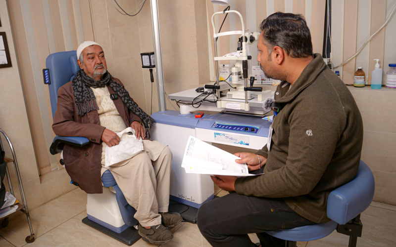
Appropriate glaucoma therapy begins with a detailed eye examination. Your ophthalmologist assesses eye pressure, optic nerve health, visual function, and the drainage angle.
Tonometry
Measures the intraocular pressure (IOP) inside the eye using precision Goldmann applanation or non-contact methods to assess baseline pressure.
Optic Nerve Examination
Checks the appearance, color, margin, and cup-to-disc ratio of the optic nerve head to detect characteristic glaucomatous cupping.
Visual Field Test (Perimetry)
Maps peripheral vision sensitivity to detect subtle blind spots (scotomas) and assess the functional extent of any vision changes.
OCT Scan (Retinal Nerve Fiber Layer)
High-resolution optical coherence tomography measures microscopic thickness of the retinal nerve fiber layer (RNFL) and ganglion cell complex.
Gonioscopy
A specialized mirrored lens evaluates the anatomical angle where the iris meets the cornea to distinguish open-angle from angle-closure glaucoma.
Clinical Interpretation & Protocol
Together, these comprehensive tests establish diagnosis, stage severity, define individualized target IOP, and guide long-term monitoring.
Best Glaucoma Treatment in Lucknow: Available Treatment Options
Controlling eye pressure is the primary aim of treating glaucoma and slowing optic nerve damage. Treatment is usually lifelong and tailored to each patient.
01. Glaucoma Eye Drops
Medicated drops work by reducing aqueous fluid production or improving fluid outflow through the eye's natural drainage channels.
Explore Medications02. Laser Treatment
Advanced laser procedures like SLT and LPI improve natural fluid outflow or relieve pupil block, often reducing medication dependency.
Explore Laser Options03. Glaucoma Surgery
Surgical procedures such as trabeculectomy, tube shunt drainage devices, or MIGS create new drainage pathways when drops or laser are insufficient.
Explore Surgical CareGlaucoma Eye Drops
For many patients, glaucoma eye drops are the first-line treatment for controlling elevated eye pressure. These medicines work by reducing fluid production or improving fluid drainage.
Prostaglandin Analogues
Increase fluid outflow through the secondary uveoscleral pathway; typically administered once daily at bedtime.
Beta Blockers
Work by decreasing the rate of aqueous humor fluid production within the ciliary body.
Alpha Agonists
Provide dual action: reduce fluid production while simultaneously improving uveoscleral drainage.
Carbonic Anhydrase Inhibitors
Eye drops that inhibit carbonic anhydrase enzymes, reducing fluid secretion inside the eye.
Combination Drops
Combine two different medication classes into a single bottle for enhanced convenience and adherence.
Important Medication Guidance
Benefits: Non-surgical, effective for many patients, and easy to use. Adherence: Regular use is important because missing prescribed doses may allow eye pressure to rise. Therefore, patients should use their medication exactly as directed by their ophthalmologist and attend scheduled follow-up appointments.
Laser Treatment for Glaucoma in Lucknow
Laser treatment can help lower eye pressure by improving the drainage of fluid from the eye for certain types of glaucoma.
Selective Laser Trabeculoplasty (SLT)
A gentle, targeted laser procedure that applies low-energy pulses to specific pigmented cells in the trabecular meshwork to improve natural fluid drainage.
- Common for open-angle glaucoma
- Improves natural fluid drainage
- Can reduce the daily need for eye drops
- Outpatient procedure with minimal discomfort and quick recovery
Laser Peripheral Iridotomy (LPI)
A preventive or therapeutic laser treatment that creates a microscopic opening in the outer edge of the iris to restore natural fluid circulation between eye chambers.
- Used for angle-closure glaucoma or narrow angles
- Balances pressure between anterior and posterior chambers
- Prevents sudden, dangerous pressure rises (acute attacks)
- Quick in-clinic procedure performed with topical numbing drops
When Is Glaucoma Surgery Considered?
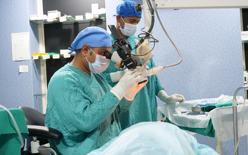
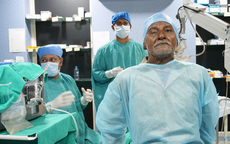
If medicines or laser treatment do not provide sufficient control, glaucoma surgery may be recommended. The appropriate procedure depends on the type of glaucoma, severity, optic nerve condition, and individual needs.
Trabeculectomy
Creates an alternative microscopic drainage flap under the conjunctiva (filtering bleb), allowing excess aqueous humor to bypass clogged drainage channels and lower intraocular pressure.
- Creates alternative fluid outflow pathway
- Proven gold standard for progressive glaucoma
- Close postoperative monitoring ensures optimal bleb function
Drainage Devices & MIGS
Glaucoma drainage devices (tube shunts) can be considered in advanced or complicated cases. Selected patients with mild to moderate glaucoma may also be suitable candidates for minimally invasive glaucoma surgery (MIGS).
- Drainage Devices: Tiny silicone tube implants for refractory cases
- MIGS: Microscopic stents with favorable safety profile and rapid recovery
- Goal: Controls eye pressure to preserve remaining vision (does not restore lost nerve tissue)
Glaucoma Treatment Cost in Lucknow
Glaucoma care is customized to each patient’s clinical stage. Treatment costs depend on several individualized factors rather than a single fixed figure.
Type & Stage of Glaucoma
Early-stage monitoring differs in protocol from advanced or complicated cases requiring intensive pressure control.
Diagnostic Tests Required
Specialized tests such as high-definition OCT RNFL scans, visual field perimetry, and pachymetry corneal thickness measurements.
Medications Prescribed
Specific classes of prescription eye drops, whether monotherapy or multi-agent combination formulations are required.
Laser or Surgery Needed
Whether outpatient laser therapy (SLT/LPI) or microsurgical intervention (trabeculectomy, shunt device, MIGS) is indicated.
Hospital Infrastructure
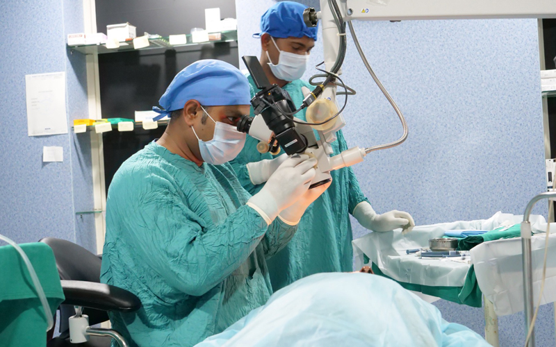
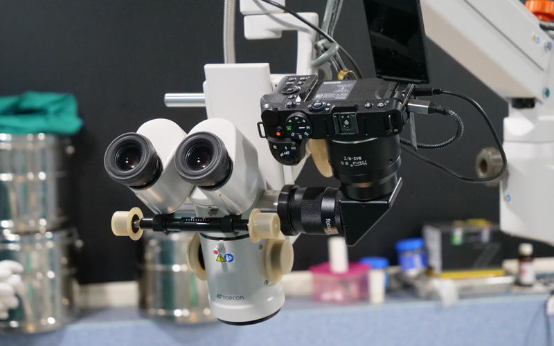
Advanced ophthalmic diagnostic suites, specialized surgical microscopes, and NABH-accredited sterile clinical environments.
Doctor’s Specialized Expertise
Comprehensive evaluations and surgical interventions performed by fellowship-trained glaucoma and ophthalmic specialists.
Long-Term Follow-Up After Glaucoma Treatment
Glaucoma is a lifelong condition, and the disease can sometimes progress even when eye pressure is being treated. Regular follow-up appointments are essential for protecting remaining vision.
Routine Visit
Regular scheduled clinical checkups.
Check Eye Pressure
Accurate IOP tonometry reading.
Repeat Field & OCT
Compare serial scans with baselines.
Adjust Drops
Modify medication if IOP fluctuates.
Detect Progression
Identify subtle structural nerve changes.
Evaluate Care
Verify effectiveness of treatment.
Glaucoma and Cataract Treatment
Cataracts and glaucoma frequently coexist in older adults. Comprehensive evaluation determines whether sequential or combined management is most appropriate.
-
Pressure Reduction from Cataract Surgery: Just the cataract surgery itself can bring intraocular pressure down a bit by deepening the anterior chamber.
-
Simultaneous Surgical Approaches: Simultaneous cataract and glaucoma surgery (phaco-trabeculectomy) may be recommended in selected clinical situations.
-
MIGS in Combination: Minimally invasive glaucoma surgery (MIGS) micro-stents can often be placed through the same small cataract incision.
Good planning addresses visual clarity and eye-pressure management simultaneously, minimizing operative interventions while optimizing long-term visual prognosis.
Who Needs Glaucoma Screening and Treatment?
High-risk persons should undergo periodic glaucoma screening. Because early glaucoma is silent, screening is the most reliable way to catch changes before vision is lost.
Over 40 Years Old
Risk increases progressively with age.
Family History
Having glaucoma incidence in parents or siblings.
Diabetes & High BP
Systemic conditions affecting ocular circulation.
Long-Term Steroid Use
Oral, topical drops, or inhaler steroid medicines.
High Eye Pressure
Previously noted elevated intraocular pressure.
Previous Eye Injury or Surgery
Past blunt ocular trauma or prior intraocular surgery.
Trusted Glaucoma Treatment in Lucknow
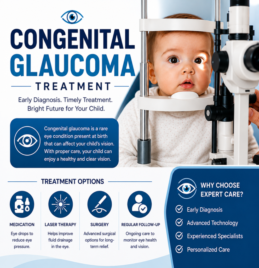
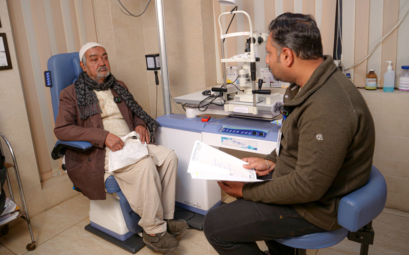
For patients in search of trusted and progressive glaucoma evaluation and care, Klarity Eye Care Hospital provides complete glaucoma diagnosis, medical therapy, laser therapy, and surgical treatment under the guidance of an expert specialist.
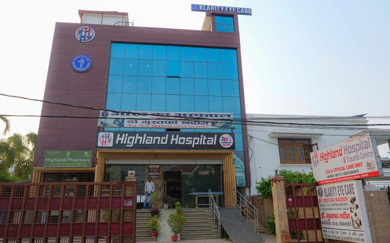
Klarity Eye Care and Multispeciality Clinic (NABH Accredited) is a leading healthcare facility in Lucknow equipped with advanced ophthalmic perimetry, optical coherence tomography, and dedicated laser suites.
NABH Accredited Hospital
B-39/1, Nirala Nagar Opp Vivekanand Hospital Lucknow

Take the Next Step Toward Better Glaucoma Care
Schedule a comprehensive glaucoma evaluation with our ophthalmic specialists at Klarity Eye Care Hospital in Nirala Nagar, Lucknow.
Request Glaucoma Consultation
Fill out the form below. Our clinical care team will contact you to confirm a convenient appointment.